Case Report

Hair dye imitating multiple basal cell carcinoma of the scalp: A misdiagnosis prevented by dermoscopy
1Eldad Silberstein, 1Vasileios A Pagkalos, 1Arsinoi A Xanthinaki, 2Oshra Sapir, 1Ofer Arnon
- 1Center of R&D in plastic surgery, Soroka University Medical Center, Ben-Gurion University, P.O.B. 151, Beer-Sheva, Israel
- 2Department of Chemical Engineering, Sami Shamoon College of Engineering, Beer-Sheva, Israel
- Submitted: April 14, 2013;
- Accepted: May 19, 2013;
- Published: May 21, 2013
This is an Open Access article distributed under the terms of the Creative Commons Attribution License (http://creativecommons.org/licenses/by/3.0), which permits unrestricted use, distribution, and reproduction in any medium, provided the original work is properly cited.
Abstract
Basal cell carcinoma is the most common malignant skin tumor. Multiple basal cell carcinomas are frequently developed in patients receiving ionizing radiation at a younger age. We present a case of multiple pigmented lesions on the scalp of a 65-years-old female with a history of radiotherapy for the treatment of tinea capitis at her childhood and a histologically confirmed basal cell carcinoma 2 years ago. The use of dermatoscopy prevented us from a misdiagnosis. This case of hair dye imitating pigmented BCC in a patient specifically prone to this type of tumor should be in mind of any plastic surgeon or dermatologist before scheduling a patient for surgery so one can avoid the stress and unpleasantness of unnecessary surgery.
Key words
Basal cell carcinoma, Skin Cancer, Tinea capitis, Radiation therapy, Dermoscopy.
Introduction
Basal cell carcinoma (BCC) is the most common malignant skin tumor [1]. BCCs have a relatively low metastatic rate and slow growth but can cause local tissue destruction and disfigurement if not early detected and treated. While treating a BCC is usually straightforward, treating multiple BCC (mBCC) is more demanding. Several factors have been suggested as risk factors of mBCC development such as old age, male sex, skin prototype (I, II), frequent sun exposure and sunburn, severe actinic damage, family history of skin tumors, low DNA repair capacity, glutathione S-transferees and cytochrome P450 polymorphism, PTCH gene polymorphism and history of radiotherapy [2-4].
Although radiotherapy is mainly used for the treatment of cancer; it has been widely used, in the past, for the treatment of non-malignant conditions as well. Today, the use of radiotherapy in non-malignant conditions is limited due to the risk of radiation-induced cancers. Patients receiving scalp radiotherapy in childhood are at increased risk of developing skin cancer with basal cell carcinoma (BCC) and squamous cell carcinoma (SCC) to be the most concerning late effects of radiotherapy [5]. Multiple BCCs often develop in patients who received ionized radiation decades before [6]. Shore et al (2002) reported a relative risk (RR) of 3.6 for BCC of the head and neck among irradiated Caucasians in response to a scalp dose of about 4.8 Gy. In their study, approximately 40% of previously irradiated patients later developed multiple BCCs, especially pigmented BCCs. Children irradiated at young age had the highest BCC risk [7]. Ron et al (1991) reported that BCC may occur subsequent to low or moderate radiation doses, as those used in the treatment of non-malignant diseases [8]. Furthermore, BCC in irradiated scalp has a more aggressive behavior and may consequently require a more aggressive surgical resection, while, there is need for a closer follow-up, as the tumor recurrence chance is higher [9].
Tinea capitis is a superficial fungal infection (dermatophytosis) of the scalp, caused predominantly by Trichophyton or Microsporum species. Although, nowadays, the treatment of choice is oral administration of griseofulvin or other antifungal drugs, in the 1930s and 1940s, tinea capitis was treated with ionizing radiation with satisfactory results [10, 11].
In this case report, we treated a patient with a history of radiation therapy for tinea capitis in her childhood and a pigmented BCC excision two years ago. The patient was referred to our department with clinical manifestations compatible with those of mBCC.
Case Report
A 65 year old female patient was referred to our department two years ago for the treatment of a pigmented lesion on her scalp. The patient had a history of scalp radiotherapy for tinea capitis during childhood. The lesion was excised and the defect was skin grafted. The histologic examination confirmed the diagnosis of BCC. The patient was put in follow-up routine.
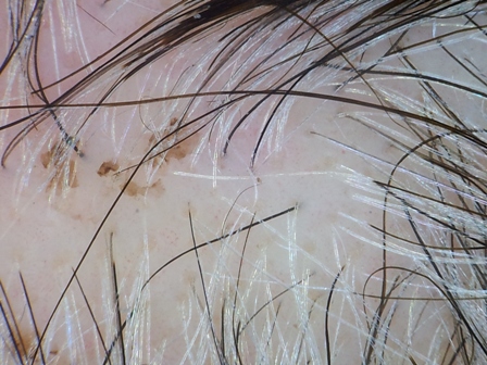
Figure 1: Pigmented lesions on the scalp
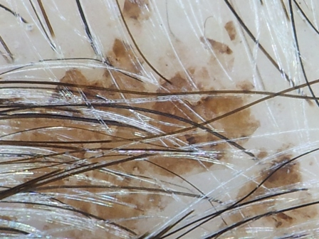
Picture 2: Dermatoscopy image of a lesion. The borders are not as irregular as initially thought
On her scheduled follow-up, the patient presented with a number of small pigmented lesions at the borders of the scar of the reconstructed area. A more thorough examination of her scalp revealed multiple pigmented lesions. The lesions were dark, multicolored with irregular borders and a diameter varying from two to fifteen millimeters (Figure 1). Taking under consideration the history of ionizing radiation at a young age and the previous diagnosis of BCC, we immediately assumed that the patient had developed mBCCs on her scalp. Dermoscopy, however, revealed that the borders of the lesions were not as irregular as they appeared to be on clinical examination (Figure 2). The lesions seemed to have a smooth, uncomplicated pattern of non-biological color material. None of the dermatoscopic criteria for BCC were present. Rubbing the lesions with a small pad leaded to the removal of the lesions (Figure 3 and 4). The patient confirmed that she had dyed her hair two weeks earlier.
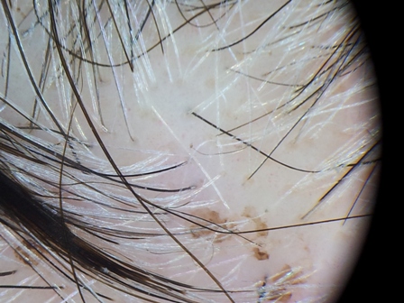
Picture 3: The lesions after rubbing the area with a small pad
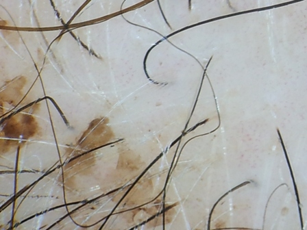
Picture 4: The lesions after rubbing the area with a small pad
Discussion
Non-melanoma skin cancers are the most common neoplasms with a growing increase in the last decades [12]. BCCsrepresentthe majority ofthesetumors [13]. Although the occurrence of multiple lesions is not uncommon, BCC usually occurs as a single lesion. Multiple BCCs may be associated with arsenic intake, burn scar, xeroderma pigmentosa, Bazex syndrome, nevoid basal cell epithelioma syndrome (Gorlin syndrome), albinism and history of ionizing radiation in the affected area [6, 8, 9, 14-16].
There are several treatment options for multiple BCCs. Although less invasive approaches may be suitable for patients of advanced age, poor general health or low risk lesions, in BCCs following radiotherapy the lesions are more aggressive and therefore require more aggressive treatment. Multiple excisions would be the treatment of choice in the case presented. The unique anatomy of the scalp and the amount of lesions make closing the excisional defects a difficult task. Skin grafting often fails and complex flaps or the importation of vascularized tissue may be required [17]. Latissimus dorsi flap is an excellent reconstructive option since it is thin, has a large surface and a long pedicle that allows it to reach all scalp regions [18].
The diagnosis of BCC is based on patient’s history, clinical examination and dermoscopic criteria presented. BCC patients often report a slowly enlarging lesion that does not heal and bleeds when traumatized. Since many clinical morphologies of BCC exist, clinical diagnosis is depended on the clinician being aware of the many forms BCC may take. Characteristic features of BCC tumors include: waxy papules with central depression, pearly appearance, erosion or ulceration, bleeding, crusting, raised borders, translucency and telangiectases. BCCs should differentialy diagnosed from a number of diseases, such as actinic keratosis, angiofibroma, Bowen’s disease, fibrous papule of the face, malignant melanoma, melanocytic nevi, psoriasis, sebaceous hyperplasia and SCC. Clinical diagnosis of BCC is very much depended on clinician’s experience and skills and can therfore often lead to missdiagnosis. Dermoscopy, on the other hand, provides the clinician with robust diagnostic criteria. Classic BCCs dermoscopic criteria include arborizing telangiectasia, blue/gray ovoid nests, ulceration, multiple blue/gray globules, leaf like areas and spoke-wheel areas. However, a number of less common dermatoscopic features can also be present in BCC lesions, such as short fine telangiectasia and multiple erosions. Even features that are suggestive of melanocytic lesions, such as multiple brown to black dots/globules, blue/white veil like structures and nonarborizing vessels can also be found in histologically verified BCC lesions. Altamura et al (2009) presented a retrospective study of the dermoscopic criteria of BBC and came to the conclusion that arborizing telangiectasia, leaf like areas and large blue/gray ovoid nests represent reliable and robust diagnostic parameters [19].
The treatment of mBCCs is demanding and therefore a misleading diagnosis such as in the case presented can be devastating. Apart from obvious consequences for the patient, the ethical, financial and legal implications of a malpractice case for the physician and the hospital should also be considered. Medical misdiagnosis occurs daily, evidenced by the high prevalence of false positive, false negative and equivocal results of diagnostic procedures. A case of misdiagnosis, however can be a significant contributor to optimize health outcomes by emphasizing the use of simple, cost-effective and fast diagnostic tools, such as, in this case, dermoscopy. None of the dermoscopic criteria, however, were present in our case.
Typically, post radiation scalp BCCs are of the pigmented type [5]. Dyeing hair is a common practice among middle aged people. This case of hair dye imitating pigmented BCC in a patient specifically prone to this type of tumor should be in mind of the plastic surgeon or dermatologist before scheduling a patient for surgery in order to avoid the stress and unpleasantness of an unnecessary surgery.
Conclusion
A well-documented predisposition, especially when the diagnosis is confirmed by histological examination a couple of years ago can easily mislead the physician. The thorough examination and the use of basic diagnostic tools should never be neglected.
Authors' Contribution
ES edited the final manuscript
VAP carried out the literature research and prepared the draft manuscript
AAX carried out the literature research and prepared the draft manuscript
OS edited the final manuscript
OF edited the final manuscript
Conflict of Interests
None
Ethical Considerations
Written consent was obtained from the patient for publication of this case report
Funding
None
Acknowledgement
None
References
[1]. Lear W, Dahlke E, Murray CA. Basal cell carcinoma: review of epidemiology, pathogenesis, and associated risk factors. J Cutan Med Surg, 2007 11(1): 19-30. [Pubmed].
[2]. van Iersel CA, et al. Prognostic factors for a subsequent basal cell carcinoma: implications for follow-up. Br J Dermatol, 2005. 153(5): 1078-80. [Pubmed].
[3]. Levi F, et al. High incidence of second basal cell skin cancers. Int J Cancer, 2006 119(6): 1505-7. [Pubmed].
[4]. Chen J, et al. Nonmelanoma skin cancer and risk for subsequent malignancy. J Natl Cancer Inst, 2008 100(17): 1215-22. [Pubmed].
[5]. Maalej M, et al., Radio-induced malignancies of the scalp about 98 patients with 150 lesions and literature review. Cancer Radiother, 2004. 8(2): 81-7. [Pubmed].
[6]. Boaventura P, et al., Head and neck basal cell carcinoma prevalence in individuals submitted to childhood X-ray epilation for tinea capitis treatment. Eur J Dermatol, 2012; 22(2): 225-30. [Pubmed].
[7]. Shore RE, et al., Skin cancer after X-ray treatment for scalp ringworm. Radiat Res, 2002 157(4): 410-8. [Pubmed].
[8]. Ron E, et al., Radiation-induced skin carcinomas of the head and neck. Radiat Res, 1991 125(3): 318-25. [Pubmed].
[9]. Hassanpour SE, et al., Basal cell carcinoma of scalp in patients with history of childhood therapeutic radiation: a retrospective study and comparison to nonirradiated patients. Ann Plast Surg, 2006. 57(5): 509-12. [Pubmed].
[10]. Gupta AK, Summerbell RC. Tinea capitis. Med Mycol, 2000. 38(4): 255–87. [Pubmed].
[11]. Gupta AK, Cooper EA, Update in antifungal therapy of dermatophytosis. Mycopathologia 2008. 166 (5-6): 353–67. [Pubmed].
[12]. Kopke, L. and S. Schimidt, Carcinoma basocelular. An Bras Dermatol, 2002. 77: p. 249-82.
[13]. Dahl E, et al., Basal cell carcinoma. An epidemiologic study in a defined population. Cancer, 1992 70(1): 104-8. [Pubmed].
[14]. Wagner SL, et al., Skin cancer and arsenical intoxication from well water. Arch Dermatol, 1979 115(10): p. 1205-7. [Pubmed].
[15]. Margolis MH. Superficial multicentric basal cell epithelioma arising in thermal burn scar. Arch Dermatol, 1970 102(4): 474-6. [Pubmed].
[16]. Southwick GJ, Schwartz RA. The basal cell nevus syndrome: disasters occurring among a series of 36 patients. Cancer, 1979 44(6): 2294-305. [Pubmed].
[17]. Hussussian, C. and G. Reece, Microsurgical scalp reconstruction in the patient with cancer. Plast Reconstr Surg, 2002 109(6): 1828-34. [Pubmed].
[18]. Nasir S, et al., Coverage of the cranium with latissimus dorsi in the recurrent basal cell carcinoma of Gorlin syndrome: for protection against clinical invasion. J Craniofac Surg, 2006 17(3): 599-602. [Pubmed].
[19]. Altamura D, et al., Dermatoscopy of basal cell carcinoma: morphologic variability of global and local features and accuracy of diagnosis. J Am Acad Dermatol, 2010 62(1): 67-75. [Pubmed].

