Case Report

A Giant Primary Supraumbilical Hernia Coexisting With A Giant Lipoma: A Case Report.
1Jerry Godfrey Makama, 2Stephen Ekundayo Garba
- Submitted: December 26, 2013
- Accepted: February 12, 2014
- Published: March 16, 2014
This is an Open Access article distributed under the terms of
the Creative Commons Attribution License
(http://creativecommons.org/licenses/by/3.0)which permits unrestricted use, distribution, and reproduction in any medium, provided the original work is properly cited
Abstract
Background
A ventral hernia is a hernia involving the anterior abdominal wall. It is further divided into primary abdominal wall hernias (those hernias that occur spontaneously in the anterior abdominal wall with no previous history of abdominal wall surgery or trauma) from incisional hernias (occurring from a weak scar in the anterior abdominal wall). Giant ventral hernias are considered in cases where the hernia orifice is greater than 10 cm. Giant lipoma are considered when weight is < 5kg.
Case Presentation
A 30 year-old female was admitted in the surgical ward with a three year history of progressive, painless abdominal swelling above the umbilicus. The swelling does not disappear, especially, when lying supine. Examination revealed an obese woman, with a BMI of 40.1, well hydrated and pink. Her temperature was 36.8 ºC. The cardiopulmonary status was normal with a pulse rate of 89/min, blood pressure of 110/78 mm Hg and the lung fields were clear. There was a swelling around the umbilicus with no visible peristalsis; no visible and palpable cough impulse. The swelling was soft with no area of tenderness; however, it was difficult to reduce. Digital rectal examination was normal. Haemogram was 10.1g/dl. Abdomino-pelvic ultrasound scan of the abdomen reported loculated fluid and some gas levels suggestive of a huge mass of various loops of bowel adjourning fatty tissue in lobules in the anterior abdominal wall. Intra-operatively, the abnormalities found were an invagination of loops of jejunum through a defect and fatty tissue (lipoma) in the anterior abdominal wall. Following excision of the lipoma and successful manual reduction, a wall defect of about 11.8cm was found. The defect was primarily repaired with a non absorbable Nylon suture. Patient did well postoperatively and was discharged after 10 days. The patient has been entirely free of symptoms since six months of operation.
Conclusion
Careful preoperative preparation, operative technique, and postoperative care are required for successful management of giant primary supra-umbilical hernias coexisting with a lipoma
Key words
Giant, Hernia, Lipoma, Ventral
Introduction
Since 2000, several authors have proposed further classifications for abdominal hernias, particularly ventral hernias [1, 2, 3]. Attempts have been made to distinguish ventral hernias into primary abdominal wall hernias (those hernias that occur spontaneously in the anterior abdominal wall with no previous history of abdominal wall surgery or trauma) from incisional hernias, (occurring from a weak scar in the anterior abdominal wall). Giant ventral hernias are considered in cases where the hernia orifice is greater than 10 cm [2, 3, 4]. Giant hernias are more liable to complications and poorly controlled by external support. The management of these giant hernias has been a major challenge to the surgical community with various suggestions and options ranging from simply repair to components separation and use of prosthesis [2, 3, 5]. These options have resulted from a basic fact that giant hernias are usually associated with loss of abdominal realm which occurs when the intra-abdominal contents can no longer lie within the abdominal cavity [1, 4].
There are many problems associated with the management of such giant hernias. We therefore wish to report a case of giant primary supra-umbilical hernia coexisting with a giant lipoma and also, to review the literature.
Case Report
A 30 year-old female Nigerian was admitted in the surgical ward with a three year history of progressive, painless abdominal swelling above the umbilicus. At the beginning, it occasionally disappeared, especially, when lying supine. . However, in the previous 6 months it was increasing in size so rapidly, to the extent that, it was now compromising her mobility, associated with dragging sensation on the anterior abdominal wall and was not disappearing on lying down any longer even if attempt were made to reduce manually. All along pregnancy test has been negative. No history of upper gastrointestinal endoscopy or imaging studies. No history of previous abdominal surgery or trauma, no cardio-respiratory or Genito-urinary symptoms, no obvious referable etiological factor. However, she admitted been obese in the last one year.
Physical examination revealed an obese woman with a BMI of 40.1, who was well hydrated and pink. She was afebrile with a temperature of 36.8°C. The cardiopulmonary status was normal with a pulse rate of 89/min, blood pressure of 110/78mmHg and lung fields were clear. There was a swelling around the umbilicus (Figure 1), no visible peristalsis; no visible and palpable cough impulse. The swelling was soft with no area of tenderness; no spontaneous reduction on lying supine and on manual reduction. Digital rectal examination was normal.
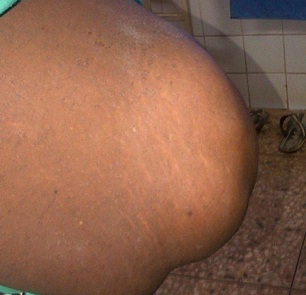
Figure 1: Herniation around the Umbilicus
Haemogram was 10.1g/dl. Abdomino-pelvic ultrasound scan of the abdomen reported loculated fluid and some gas levels suggestive of a huge mass of various loops of bowel adjourning lobulated fatty tissue. After a five day pre-operative bowel preparation, she consented to surgery. The giant mass was approached through a midline incision. Intra-operatively the findings were that of a giant lipomatous tissue and an invagination of the terminal portion of the jejunum and then the ileum and pre-peritoneal fat tissue (Figure 2) into the anterior abdominal wall. Further exploration revealed a giant mass of fatty tissue attached to herniated bowel (Jejunum and omentum) (Figure 3) bowel, including pre-peritoneal fat was found to be viable and thus was manually reduced. Following successful manual reduction, a wall defect of about 11.8cm (Figure 4) was found. Lateral release incisions were made bilaterally on the external aponeurosis. A new midline was formed with continuous suture taken from the medial edge of one released incision to the one on the other side. Thereafter this new midline was re-enforced with the fascia (Figure 5) and, following removal of excess skin, abdomen was closed using non absorbable Nylon 2/0. The giant lipomatous mass (Figure 6) was, however, sent for histology which revealed normal adipocytes. Patient did well postoperatively and was discharged after 10 days. The patient has been entirely free of symptoms since six months of operation.
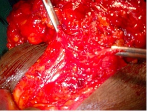
Fig.2. Invagination of the contents of the hernia through the defect
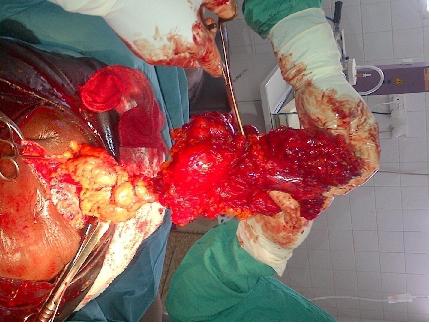
Fig.3. Characteristics of the contents of the hernia (From top down-Lipoma, Omentum and bowel)
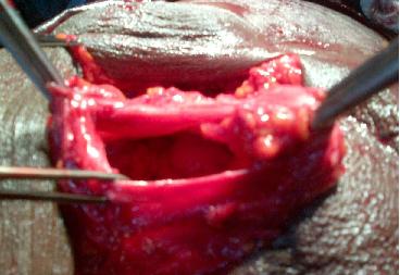
Figure 4 The Anterior abdominal wall defect
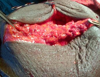
Fig.5. After repair of defect
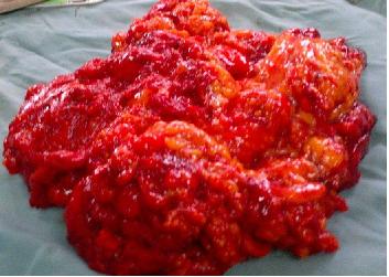
Figure 6. The Lipomatous tissue after excision (Weight=5.6 Kg)
Discussion
Giant primary supra-umbilical hernias are uncommon. The width of the hernia defect has been the major and only one parameter to classify giant hernias, also, stating that the width is the most important measurement of size to determine the difficulty of successfully repairing the hernia [1, 2, 7]. They present a new spectrum of problems for patients apart from the classical complications of surgical correction of defect [6, 7]. It is extremely important for surgeons to note, before patients are taken to the operating theater, that giant hernia, generally, present formidable surgical problems. Preoperatively, hernias increase gradually in size, unsightly, and are liable for severe complications. Also, patients encounter difficulties in walking, sitting or simply lying down, and their mobility is dramatically restricted [2, 4, 6, 7]. In a female patient, particularly of reproductive age, the renewed hope of been pregnant is an additional problem that may be faced by the patient. This was incredibly foremost in our case, further prolonging the need to seek surgical attention.
Giant hernias are, pre-operatively, more liable to complications and poorly controlled by external support. In giant hernias, a loss of abdominal domain occurs when the intra-abdominal contents can no longer lie within the abdominal cavity. This has been a major concern among surgeons with the popular opinion that reduction of the contents is usually difficult.
In this case it was not only a giant supraumbilical hernia but this was coexisting with a giant lipoma of the anterior abdominal wall. To our knowledge, there have been no cases in the literature of a giant supraumbilical hernia coexisting with lipoma in the anterior abdominal wall. Previous studies [8,9], have reported intra-abdominal lipoma and a hernial sac containing the uterus, ovary, and fallopian tube. Turk
et al., [8] reported a case of giant omental lipoma in an inguinal hernia sac containing the ovary, fallopian tube and the ovary. Similarly, Ozkan
et al., [9] have reported a case of a right sliding indirect inguinal hernia containing paraovarian cyst, fallopian tube and ovary. It is of utmost importance to keep in mind that hernia sac may contain almost any abdominal organ, and surgical dissection should be carried out carefully and accordingly. Pathophysiologically, these organs such as ovary, fallopian tube might be simply pulled along with contents of a hernia (sliding hernia). For this case, it is quite difficult to explain how the coexistence of this giant supraumbilical hernia with a giant lipoma in the anterior abdominal wall had happened. However, several possibilities may be postulated and entertained to this regards. It may be possible it was a serosal lipoma that protruded through the defect together with the bowel and remains quiescent for the initial time. After sometime, the lipoma may have suddenly started growing rapidly to expand into the musculo-aponeurotic planes up to its present size. On the other hand, the anterior abdominal lipoma may have become aggressive and infiltrative into the abdominal cavity to get attach to the loops of bowel. However, the lipoma was well encapsulated intraoperatively, with histology of benign adipocytes. There seems to be a need for clinical and experimental studies to further explain the mechanisms that apply to the pathogenesis of this type of coexistence.
Morbidity and mortality associated with the repair of giant hernia have been considered high [10, 12]. This is largely due to the fact that the abdominal cavity (already contracted) due to longstanding of the abdominal content outside its domain may compromise cardio-respiratory status when returned into the abdominal cavity. Hence, postoperative disorders in the cardiovascular system, inadequate tissue oxygenation, increased intra–abdominal pressure, and pulmonary embolism may further expose the patient to severe risks [6, 8]. Also, it is known that hernias that are large, the risk of recurrence is usually high [8]. Lastly the residual skin usually needs excision for cosmetic reasons.
It has been agreed and advised that all hernias, particularly the massive one, should be repaired unless the patient is unable or unwilling to undergo surgery [7, 14, 15]. This informed our desire to repair this index case as soon as theater space could permit.
Adequate pre-operative preparation has been shown to reduce significantly the morbidity and mortality [13] associated with this clinical problem. In this case, a five day bowel preparation was done using low residue diet, liquid diet, and colonic lavage. It has been established that aqueous sodium phosphate is a hyper-osmotic solution and often draw plasma into the bowel lumen, thus, promote colonic emptying and cleansing. This is thought to, reliably, empty the colon of fecal material, not cause any patient discomfort or harm and would reduce intra-abdominal pressure [13]. Haven ensured a roamy abdominal cavity from above measures, attempt to close the musculo-aponeurotic defect went on and was done with ease. This was done in other to cut cost as the patient could not afford a mesh for repair. The repair did not compromise respiration or cardiac functions. The patient did not required ventilation or suffered from compartment syndrome after operation and we felt, this pre-operative bowel preparation and excision of the redundant giant lipoma (contributing 60% of the total mass) contributed significantly to the smooth perioperative recovery of the patient.
Conclusions
Careful preoperative preparation, operative technique, and postoperative care are required for successful management of giant primary supra-umbilical hernias coexisting with a giant lipoma. The use of host tissue is relatively simple, safe, and a reliable surgical solution to the problem of giant supra-umbilical hernia particularly in an extremely poor patient like the index case.
Learning points
1. This is an extremely rare case of giant primary supra-umbilical hernia
2. Also giant lipoma in the anterior abdominal wall
3. The coexistence of the two will absolutely raise interest among surgeons on how it came about including other consequences (both physical and social) that may bear on the patient
Authors' Contribution
J G M conceived the study, participated in design, carried out literature search, prepared the manuscript and edited the final manuscript.
E S G participated in literature search, critical review and final approval of the manuscript.
All authors read and approved the final manuscript for submission
Acknowledgement
None
Conflict of interest
The authors declare that there are no conflict of interests
Ethical Consideration
Written informed consent was obtained from the patient for
publication of this case report.
References
[1]. Muysoms FE, Miserez M, Berrevoet F, Campanelli G,
Champault GG, Chelala E, Dietz UA, Eker HH, El Nakadi I, Hauters P, Hidalgo
Pascual M, Hoeferlin A, Klinge U, Montgomery A, Simmermacher RK, Simons MP,
Smietański M, Sommeling C, Tollens T, Vierendeels T, Kingsnorth A.
Classification of primary and incisional abdominal wall hernias. Hernia 2009;
13: 407-414 [Pubmed].
[2] Chevrel JP, Rath AM. Classification of incisional hernias of the abdominal wall. Hernia 2000; 4:7–11
[3] Flament JB and Palet JP. Prosthetic repair of massive abdominal ventral hernias. In: Nyhus and Condon’s Hernia 5th edition). Lippincott W&W, Philadelphia, USA, 2002: 341-366.
[4]. Ammar SA. Management of Giant Ventral Hernia by
Polypropylene Mesh and Host Tissue Barrier: Trial of Simplification. J Clin Med
Res • 2009; 1(4):226-229 [Pubmed].
[5] Al Sarakbi W, Agrawal A, Taffinder N. A giant
inguinoscrotal hernia: a case report and review of the literature. Grand Rounds
2005; 5: 46–48
[6] King JN, Didlake RH, Gray RE. Giant inguinal hernia.
South Med J 1986; 79(2):252-253 [Pubmed].
[7] Ammaturo C, Bassi G. The ratio between anterior abdominal wall surface/wall defect surface: a new parameter to classify abdominal incisional hernias. Hernia 2005; 9:316–321 [Pubmed].
[8]. Turk E, Karagulle E, Oguz H, Toprak E Indirect
hernial sac containing the uterus, ovary, and fallopian tube in association with
a giant intraabdominal lipoma: report of a case. Hernia. 2012 Oct;16(5):593-5 [Pubmed].
[9] Ozkan OV, Semerci E, Aslan E, Ozkan S, Dolapcioglu K, Besirov E. A right sliding indirect inguinal hernia containing paraovarian cyst, fallopian tube, and ovary: a case report. Arch Gynecol Obstet 2009 Jun;279(6):897-9. doi: 10.1007/s00404-008-0807-0
[10] Paajanen H, Laine H. Operative treatment of massive ventral hernia using polypropylene mesh: a challenge for surgeon and anaesthesiologist. Hernia. 2005; 9:62-7.
[11] Tran NV, Petty PM, Bite U, Clay RP, Johnson CH, Arnold PG. Tissue expansion-assisted closure of massive ventral hernias. J Am Coll Surg 2003;196(3):484-488 [Pubmed]
[12]. Mehendel FV, Taams KO, Kingsworth AN. Repair of a
giant inguinoscrotal hernia. British J Plastic Surgery 2000; 53: 525–9 [Pubmed].
[13]. Shawki S, Wexner SD. How safe is bowel preparation
with oral sodium phosphate solution? Nat Clin Pract Gastroenterol Hepatol
2008;5(9):482-483 [Pubmed].
[14] de Vries Reilingh TS, van Goor H, Charbon JA, Rosman
C, Hesselink EJ, van der Wilt GJ, Bleichrodt RP. Repair of Giant Midline
Abdominal Wall Hernias: ‘‘Components Separation Technique’’ versus Prosthetic
Repair. World J Surg 2007; 31: 756–763 [pubmed]
[15]. Ramirez OM, Ruas E, Dellon AL. Component separation method for closure of abdominal wall defects: an anatomic and clinic study. Plast Reconstr Surg. 1990;86:519-26[pubmed]

