Case Report

Scrotal fibroangioma: A case report
1Satyendra K Tiwary, 1Ajay K Khanna
- 1Department of General Surgery Institute of Medical Sciences, Banaras Hindu University,Varanasi 221005.
- Submitted: October 01, 2012
- Accepted October 15, 2012
- Published: October 1, 2012
This is an Open Access article distributed under the terms of the Creative Commons Attribution License (http://creativecommons.org/licenses/by/3.0), which permits unrestricted use, distribution, and reproduction in any medium, provided the original work is properly cited.
Introduction
Fibroangioma is a rare benign tumor and is located most commonly in nasopharynx but it has been reported to occur in different parts of the body. No case of fibroangioma scrotum has been reported in the literature.
Case report
We present a patient of scrotal fibroangioma and our experience in managing it. A 48 year male presented with recurrent discharge of blood from scrotum for last three months. We did partial scrotectomy to excise the involved skin and scrotoplasty to close the wound. Post-operative recovery was uneventful with patient becoming symptom free in follow-up period.
Conclusions
Scrotal fibroangioma should be managed on the same principle of its management elsewhere in the body. Painless bleeding from scrotum without fever or itching has high chances of being fibroangioma.
Keywords
Partial scrotectomy, Scrotoplasty, Scrotum, Fibroangioma, Tumor, Benign.
Introduction
Fibroangioma is a rare tumor of the body. Though it has been reported in literature to originate in different body parts, so far no case of scrotal fibroangioma has been reported. We present a case of fibroangioma scrotum with frequent blood discharge from scrotum which was completely cured with partial scrotectomy and scrotoplasty.
Case Report
A forty eight years old male was referred from dermatology outpatient department to surgical outpatient department with painless discharge of blood from scrotum since last three months. He has taken medications advised by the dermatolgist, but no improvement was noticed. The patient gave history of incision and drainage for scrotal abscess six years back. After that he had one or two episodes of blood discharge from scrotum every year. But for last three months, he had frequent painless discharge of blood from scrotum without fever and itching.
On clinical examination, maculo-papular eruption localized in central two third of scrotum was noted (Figure 1). On squeezing scrotal skin, blood tinged discharge was present. Corrugations of scrotal skin were present. No thickening of scrotal skin was noted. Inguinal lymph nodes were enlarged on both sides with sizes of 0.5cm to 1.5 cm. Testis, spermatic cords, penile skin, external urethral meatus were normal on clinical examination.
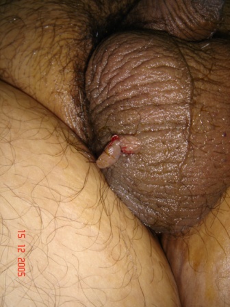
Figure 1. Clinical photograph of fibroangioma scrotum.
Wide local excision was planned. Under local anesthesia, elliptical incision encircling lesion was given with a minimum margin of 0.5 cm. Partial scrotectomy was done and scrotoplasty was performed. Gross examination of excised tissue revealed marked vascularity of the tissue underlying the skin. Microscopic examination showed fibroangiomatous tissue covered by skin (Figure 2). Histopathological diagnosis was fibroangioma scrotum. Postoperative recovery was uneventful with slight serous discharge through incision. On day ten, stitches were removed. There was no discharge or any other complaint. Patient is doing well at follow-up of six months.

Figure 2. Microphotograph of fibroangioma scrotum.
Discussion
Fibroangioma is a rare benign tumor of body. It is a hamartoma comprising of vascular and fibrous elements. There may be many triggering factors for development of fibroangioma from dormant nidus. Most well established triggering factor is testosterone as it is a disease of male predominantly in second decade of life. Commonest site of fibroangioma is nasopharynx. Other sites of fibroangioma reported in literature are-tongue [1], larynx, spermatic cord [2], buttocks, ileum, uterus, ureter [3], urethra, cervix and digits [4]. Fibroangioma of scrotum has not been reported in literature. Vascular and fibrous elements may vary in composition. Vascular elements are deficient in muscular components and only endothelial cells are present. Due to absent muscular components, blood vessels do not contract leading to troublesome bleeding which is predominant presentation in fibroangioma. Causes of bleeding from scrotum are usually due to tumor, infection, angiokeratoma or varices [5]. Fibroangioma may cause obstruction if occupies lumen of any organ.
Different treatment modes have been suggested but surgery remains cornerstone of all treatment modalities. Other modes of treatment are preoperative radiotherapy but not found to offer significant benefit. Cryotherapy has been used but not established. Estrogen is used to reduce bleeding. Definitive mode of treatment is excision of the lesion. Recurrence has also been reported after excision.
Conclusions
Scrotal fibroangioma should be managed on the same principle of its management elsewhere in the body. Painless bleeding from scrotum without fever or itching has high chances of being fibroangioma. Surgical excision of lesion comprising partial scrotectomy and scrotoplasty should be the treatment of choice. If skin loss is extensive and primary closure is not possible then either skin grafting or placing testis in thigh pouches or abdominal pouches may be done. Recurrence of fibroangioma of other sites has been reported, so scrotal fibroangioma also has recurrence potential.
Authors’ contribution
SKT: Did the literature search and prepared the draft manuscript.
AKK: Concept and design, editing the final version for publication.
Acknowledgement
None
Funding
None
Ethical Considerations
A written informed consent was obtained from the patient for publication of this case report.
References
[1]. Picarelli A, Rafaelli R. A case of fibroangioma localized on the tongue occurring during pregnancy. Riv Ital Stomatol. 1977; 46: 10-13.[Pubmed]
[2]. Macellari G, Giustina A. A case of fibroangioma of spermatic cord. Minerva Urol.1980; 32: 179-82.[Pubmed]
[3]. Frattini A, Ferretti S, Peracchia G, Simonazzi M, Cortellini P. Polyfibroangioma of the ureter. Endoscopic treatment. Acta Biomed Ateneo Paramense.1995; 66: 243-7.[Pubmed]
[4]. Kohda H, Narisawa Y. Digital verrucous fibroangioma: a new variant of verrucous hemangioma. Acta Derm Venereol.1992; 72:303-4 [Pubmed]
[5]. Khanna AK, Seth P, Khanna R. Bleeding scrotal varices as presentation of Budd Chiari Syndrome. Indian J Gastro. 2000; 19: 190-1.[Pubmed]

