Case Report

Congenital Orbital Teratoma with Bilateral Anopthalmia: A Rare Presentation
Rajendra P. Maurya1, Virendra P. Singh1, Prashant Bhushan1, Mahendra K. Singh1, Mohan Kumar2
- 1Department of Ophthalmology, Institute of Medical Sciences, Banaras Hindu University, Varanasi India
- 2Department of Pathology, Institute of Medical Sciences, Banaras Hindu University, Varanasi India
- Submitted: March 27, 2012;
- Accepted April 12, 2012
- Published: April 16, 2012
This is an Open Access article distributed under the terms of the Creative Commons Attribution License (http://creativecommons.org/licenses/by/2.0), which permits unrestricted use, distribution, and reproduction in any medium, provided the original work is properly cited.
Background:
Teratomas are rare embryonal tumors that comprise approximately 1% of orbital tumors in childhood.
Case report:
This article is to describe a rare congenital orbital tumor arising in an eight and a half month old male child having bilateral anophthalmos. Complete excision of the mass was done. Histopathological examination revealed true teratoma. Follow-up after one year showed no recurrence.
Conclusions:
The pathogenesis, clinical features, histopathological findings and treatment of orbital teratoma are discussed.
Introduction
Teratomas are rare embryonal tumors that comprise approximately 1% of orbital tumors in childhood [1]. Teratomas are benign in nature and comprise of a wide variety of tissues derived from all the three germ layers (ectoderm, mesoderm and endoderm). Here an unusual case of congenital teratoma associated with bilateral anophthalmos is being presented.
Case Report
An eight and a half month old male child whose gestation and delivery records were normal, presented with a mass in right orbit, since birth. It was increasing rapidly. At the time of examination, a huge mass of variable consistency in the right orbit measuring 7 cm x 6.5 cm was present. The mass was non-pulsatile, irreducible and non-transilluminant. It was nontender and did not increase in size on coughing and crying. Skin over the mass was overstretched and tense. Eyelids and palpebral fissure could not be visualized. Periocular skin was overstretched (Figure 1). Tumor did not show any structure of the eye ball. Eye ball was absent in the left orbit since birth. General examination was within normal limit. There were no other congenital defects in the body.
Radiological examination revealed huge orbital mass causing enlargement of orbit in both dimension without bony invasion or erosion. A provisional diagnosis of orbital dermoid was made and the patient was taken up for a total excision of tumor under general anesthesia. Postoperatively patient had an uneventful recovery. The cut section of the gross specimen revealed yellowish white irregular mass of variegated consistency with large areas of multiple cysts containing fluid. Multiple histological section demonstrated derivatives of all the three germ cell layers in the form of an adipose tissue, cartilage, neuroglial cells, gastric mucous, mucous glands and cysts lined by respiratory epithelium. There was no microscopic evidence of ocular tissues within the teratoma (Figure 2). A conclusive diagnosis of mature teratoma in an anophthalmic eye was made. One year post operative follow up showed no recurrence.
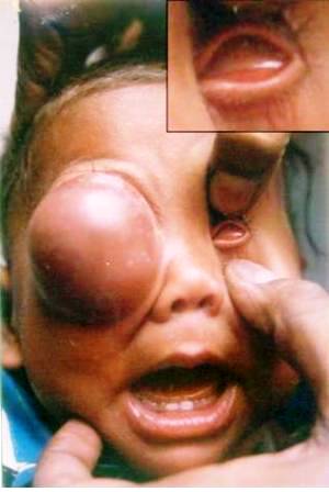
Figure 1: large cystic mass in the right orbit with anophthalmos left side (Inset: left eye showing absence of eyeball.
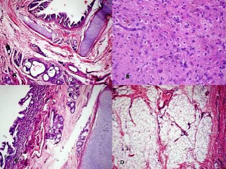
Figure 2: Photomicrograph showing A) Immature cartilage, respiratory epithelium and glandular epithelium. B) Neuroglial tissue C) Immature cartilage, Gastric mucosa and mucus glands. D) Lobules of adipose tissue.
Discussion
The word “teratoma” in Greek means monstrous growth. Orbital teratoma is a rare congenital benign tumor arising from all the three germ layers [2,3]. These totipotent cells are foreign to the orbit [4]. Teratomas are considered to be choristomas rather than true neoplasm [5]. Choristomas are foci of normal tissues located in abnormal sites. First case of congenital orbital teratoma was reported by Holms in 1862 [6] which was actually a teratoid tumor. Majority of teratomas are teratoid consisting of only ectodermal and mesodermal tissues and rarely consist of mesodermal and endodermal tissues [7,8]. Commonly occurring congenital benign orbital tumors like dermoid, angiomas and neurofibroma etc. are derived from one germ layer. The first true teratoma which arisen from three germ layers, was reported by Broer and Weigert [9]. Clinically it is very difficult to distinguish orbital teratoma with other congenital benign orbital tumors [10]. Orbital dermoid and microopthalmos with cystic eye are common differential diagnosis. In the present case, eye ball could not be seen in teratomatous orbit and ocular contents could not be identified even in histopathology which shows possible anophthalmos. Pathogenesis of our case may be an expression of a genetic abnormality as primary anophthalmos appears due to result from failure of the optic pit to deepen and form an optic out growth from the forebrain [11]. Clinically congenital orbital teratomas usually grow rapidly after birth [12], however slow growing cases have been reported in adults [13]. Orbital teratomas are benign, do not erode into bone or extend into the cranium [7,8,9]. However few authors have reported extension of benign teratoma into perioribital sinus and cranial fossa [12]. Soares et al., reported malignant teratoma of orbit [14]. Garden JW et al., reported a case of orbital teratoma with subsequent malignancy during recurrence [15].
Histopathological examination of teratoma usually show the predominant germ cell type to be ectodermal like hair follicles, sweat glands and neuroglial cells; followed by mesoderm represented by fat, muscles and bone cartilage. Endoderm is least common represented by gastrointestinal tissues or cysts lined by respiratory epithelium [10]. In our case derivatives of all the three germ layers were found.
Conclusion
Congenital orbital teratoma with bilateral anophthalmos is a rare presentation. Teratoma should be a differential diagnosis of orbital mass in an infant or children. Early surgical removal is recommended for cosmetic purposes and to prevent the chance of rupture or malignant transformation.
Authors’ Contributions
RPM: Performed literature search, carried out the study and prepared draft and manuscript.
PB: Performed literature search and conducted the study under his guidance.
VPS: Collected, interpreted and analysis of the clinical data.
MKS: Designed the study, helped with analysis and interpretation of the data. Read and reviewed the final manuscript.
MK: Collection, interpretation and evaluation of pathological data.
Acknowledgement
We are really very thankful to Dr. P.R. Sen, Senior Medical Officer, USHCC, Banaras Hindu University for drafting the manuscript and revising it critically and Dr. A.K. Verma, CMO/Ic for helping in the study.
Conflict of Interest
There are no competing interest (conflict of interest).
Ethical Considerations
The consent was obtained from the legal guardian of the patient.
References
[1]. Spinelli HM, Criscuolo GR, Tripps M, Buckley PJ. Massive Orbital teratoma in new born. Ann. Plast. Surg. 1993 Nov. 31(5) : 453-8.
[2]. Ferry AP. Teratoma of the orbit : A report of two cases. Surv. Ophthalmol. 1965; 10: 434-42.
[3]. Simonsen AH, Sogaard H. Teratoma orbitae. Report of a case. Acta Ophthalmol. Copenh. 1981; 59 : 308-316.
[4]. Assalain A, Allare G, Codere F, Polomeno RC, Brochu P, Delisle P. Congenital orbital teratoma. A clinicopathological case report including immunohistochemical staining. Can J Opthalmol. 1994; 29: 30-3.
[5]. Jensen OA. Teratoma of the orbit. Acta Ophthalmol (Copenh). 1969; 47: 317-27,
[6]. Holmes T. Congenital tumor removed from the orbit. Trans. Pathol. Soc. London 1863; 14: 248-49.
[7]. Duke-Elder S. Normal and abnormal developmental congenital deformities. In system of ophthalmology Vol. 13 Part 2 : St. Louis CV Mosby 1964: 956-97.
[8]. Hoyt, WF and Joe S. Congenital teratoid cyst of the orbit. A case report and review of the literature. Arch. Ophthalmol. 1962; 68: 196-201.
[9]. Barber JC, Barber LF, Guerry D. 3rd, Geeraets WJ. Congenital orbital teratoma. Arch. Ophthalmol. 1974; 91: 45-48.
[10]. Singh U, Subramanian A, Bal A. Congenital orbital teratoma presenting as microphthalmos with cyst. Ind J Ophthalmol 2009; 56(6) : 474-475.
[11]. Haberland C, Perou M. Primary bilateral anaphthalmia. J. Neuropathol Exp Neurol. 1969; 28: 337-51.
[12]. Weilder’ WB. Orbital teratoma removed through Kronlen incision, with a discussion of diagnosis of orbital tumors. Am. J. Ophthalmol. 1928; 11: 971.
[13]. Davis CH, Alexander E Jr. Congenital nasofrontal encephalomeningoceles and teratomas. J Neurosurg. 1959; 16: 365-77.
[14]. Soares EJC, Lopes KDS, Andreade JDS., Faleio LC, Alves JCR. Orbital malignant teratoma. A case report. Orbit 1983; 2: 235-42.
[15]. Garden JW, McManis JC. Congenital orbital-intracranial teratoma with subsequent malignancy: case report. Br J Ophthalmol. 1986; 70(2):111-3.

