Case Report

Dermatofibrosarcoma protuberans of the breast- A case report
1Deepa Rani, 1Mohan Kumar, 2Somprakas Basu, 3Amit Bhaskar
- 1Department of Pathology, 2Department of General Surgery, 3Department of Radiology, Institute of Medical Sciences Banaras Hindu University, Varanasi UP 221005, India
- Submitted: March 28, 2013
- Accepted: June 10, 2013
- Published: June 15, 2013
This is an Open Access article distributed under the terms of the Creative Commons Attribution License (http://creativecommons.org/licenses/by/3.0), which permits unrestricted use, distribution, and reproduction in any medium, provided the original work is properly cited.
Abstract
Dermatofibrosarcoma protuberans is a rare soft tissue tumour of the skin with the potential for intermediate malignancy [1]. It is a rare, locally aggressive but rarely metastasizing tumor of the deep dermis and subcutaneous tissue. It may occur at almost any site but is more common in the trunk and extremities [2, 3]. Breast is the rare site of occurrence of this entity. It is characterized by local invasion and recurrence. We report a rare case of DFSP of breast in a 14 year old girl.
AbbreviationsDFSP-dematofibrosacoma protuberance, FNAC-fine needle aspiration cytology, MRI-magenetic resonance imaging, IHC-immunohistochemistry
Introduction
Dermatofibrosarcoma protuberans (DFSP) is a rare soft-tissue neoplasm originally described in 1924 by Darier and Ferrand [4] as a progressive recurrent dermatofibroma and later named dermatofibrosarcoma protuberans by Hoffmann in 1925 [5]. It is a slow growing nodular neoplasm arising from the dermis and invading the subcutaneous tissues. It is a low to intermediate grade malignancy with a high propensity for local invasion and high recurrence rate, especially if negative resection margins are not achieved. However, metastases are rare. DFSP most commonly involves the trunk, followed by the extremities, head and neck. Occurrence in the breast is very rare [6].
Case Report
A 14 year old girl presented with a left breast lump around areola for more than a year which was gradually increasing in size. Her past medical history was insignificant apart from the lesion. On physical examination lump was about 1.5cm in size, firm and non tender. MRI findings are detailed in Figure 1.
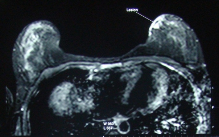
Figure 1: Figure 1: MRI of the breast showing the lesion
FNAC from the lesion showed clusters of scattered benign spindle cells and foamy histiocytes. Based on cytological finding diagnosis of DFSP was made. Lesion was then excised and on histopathological examination showed hypercellular tumour which composed of monomorphic spindle cells forming a storiform pattern throughout the dermis [figure 2].
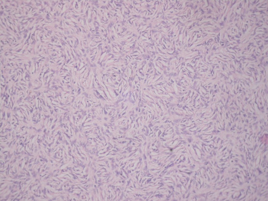
Figure 2: Figure 2: Photomicrograph showing dermatofibrosarcoma (H & E x 40)
The tumor cell showed strong immunoreactivity for CD 34 [figure 3]. The final histological diagnosis was DFSP .After 2 month of excision the patient presented with the similar lesion about 1 cm which on MRI showed heterogenous hyperintense lesion of the breast [figure 3] FNAC of that lump again showed similar appearance and diagnosis of DFSP was made.
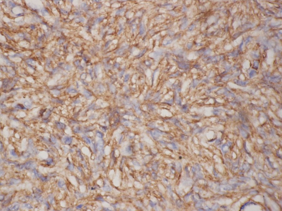
Figure 3: Photomicrograph showing immuno-histiochemistry for CD 34
Discussion
DFSP is a rare, locally aggressive soft tissue tumour arising from skin constituting approx 6%of all soft tissue sarcoma [7] with an estimated incidence of 0.8 – 0.5 cases per million per year[8].The breast is a rare site of occurrence for this uncommon entity. Only a few single case reports are found in the literature.
T.Cavusoglu et al., [1] in year 2003 reported a histologically confirmed case of DFSP in a 26 year old female. Su Ju Lee et al., [6] in the year 2009 reported the largest series of 5 cases of DFSP of breast which was diagnosed by the imaging features of DFSP. The age range was 25 -33 years. The imaging features which was included in the study were mammography , ultrasonagraphy, and MRI which defined the true subcutaneous extent of tumour.
Dimitrios M Dragoumis et al., [9] in the year 2010 reported on a 48 year old women who had a DFSP excised from her right breast and had recurrence 13 year later, diagnosis was confirmed by histopathological examination and IHC positivity of CD 34.
In our study we are reporting on a 14 year old female with early recurrence of tumour that is within 2 months of excision. Preoperatively patient was diagnosed as DFSP first by FNAC which was reported as dermatofibroma, and after excision the routine histopathology and IHC confirmed the diagnosis of DFSP. After 2 months of excision again small lump was noticed which on MRI showed heterogenous hyperintense lesion and on FNAC again came out as DFSP of breast.
Conclusion
DFSP of breast, though uncommon, does exist. With awareness of this entity, a prompt diagnosis be made and the disease be properly managed.
Authors' Contribution
DK:Prepared the draft manuscript
MKEdited the final version and contributed to pathology part of manuscript
SBHelped in preparation of manuscript, literature search
ABContributed the radiological part and prepared the manuscript
Conflict of Interests
The authors declare that there are no conflicts of interests.
Ethical Considerations
Written consent was obtained from the patient for publication of this case report.
Funding
None
References
[1]Cavusoglu T, Yavuzer R, Tuncer S. Dermatofibrosarcoma protuberance of the breast. Aesthetic Plast Surg 2003;27:104-6.[Pubmed].
[2]Fiore M, Miceli R, Mussi C, Lo Vullo S, Mariani L, Lozza L, Collini P, Olmi P, Casali PG, Gronchi A. Dermatofibrosarcoma protuberans treated at a single institution: a surgical disease with a high cure rate. J Clin Oncol 2005;23:7669-75.[Pubmed].
[3]Arnaud EJ, Perrault M, Revol M, Servant JM, Banzet P. Surgical treatment of dermatofibrosarcoma protuberans. Plast Reconstr Surg 1997;100:884-95.[Pubmed].
[4]Darier J, Ferrand M. Dermatofibromes progressifs et recidivants ou fibrosarcoma de la peau. Ann Dermatol Venereol 1924;5:545-62
[5]Hoffmann E. Ueber das knollentribende Fibrosarkom der Haunt (dermatofibrosarcoma protuberans).Dermatol Z 1925;43:1-28.
[6]Lee SJ, Mahoney MC, Shaughnessy E. Dermatofibrosarcoma of breast : Imaging feature and review of literature. AJR Am J Roentgenol 2009;193:W64-9.[Pubmed
[7]Kransdorf MJ, Meis-Kindblom JM. Dermatofibrosarcoma protuberans: radiologic appearance. AJR 1994;163:391-4.[Pubmed].
[8]Ruiz-Tovar J, Fernandez Guarino M, Reguero Callejas ME, Aguilera Velardo A, Arano Bermejo J, Cabanas Navarro L. Dermatofibrosarcoma protuberans: review of 20-years experience. ClinTransl Oncol 2006;8:606–10. [Pubmed].
[9]Dragoumis DM, Katsohi LK, Amplianitis LK, Tsiftsoglou AP. Late local recurrence of dermatofibrosarcoma in the skin of female breast. World J Surg Oncol 2010;8:48.[Pubmed].

