Case Report

Squamous Cell Carcinoma Arising in a Dentigerous Cyst- A Case Report
*Shailaja Shukla, *Mona Bargotya, *Sarika Singh, *Neha, **Pravesh
Mehra
- *Department of Pathology, **Department
of Dental Sciences, Lady Hardinge Medical College, New Delhi, India
- Submitted: Friday, February 14, 2014
- Accepted: Wednesday, February 19, 2014
- Published: Monday, March 17, 2014
This is an Open Access article distributed under the terms of the Creative Commons Attribution License (http://creativecommons.org/licenses/by/3.0), which permits unrestricted use, distribution, and reproduction in any medium, provided the original work is properly cited.
Abstract
Squamous Cell Carcinoma (SCC) arising from epithelial lining of dentigerous cyst is rare and these are distinct pathologic entity with only few cases in the literature. Primary intraosseous squamous cell carcinoma constitutes 1-2.5% of all odontogenic tumors and 1-2% of all intraoral cancers. Differential diagnosis of dentigerous cyst and the malignant tumor arising in the cyst is difficult due to nonspecific clinical and radiological examination. It is on histopathological examination that diagnosis is often made. We present a case of SCC arising from dentigerous cyst highlighting the importance of careful histopathological examination so as to prevent misdiagnosis of apparently innocuous cystic lesions.
Key words
Dentigerous cyst, odontogenic cyst, squamous cell carcinoma
Introduction
Neoplastic changes within a simple odontogenic cyst, though
rare, but are a definite entity. Neoplastic transformation among the odontogenic
cysts is highest in keratocyst and dentigerous cyst [1, 2].
Dentigerous cyst is a developmental anomaly arising during amelogenesis [3].
In rare instances, epithelial lining of the cyst can give rise to Squamous Cell
Carcinoma (SCC). Chronic inflammation and intracystic pressure are suggested as
possible casuative factors [1]. Due to nonspecific clinicoradiological findings definite diagnosis is possible by histological examination only. However, it is imperative to identify the transition from normal epithelial lining to SCC to confirm the diagnosis.
Case Summary
A 47 year old male presented with pain in lower jaw region since last four months. He gave past history of extraction of impacted bilateral canine teeth 5 months back. On oral examination, tender swelling measuring 3.5x2.5x1.5 cms at lower vestibular region with intact surrounding mucosa without any ulceration was seen.There was no associated lymphadenopathy. The panoramic X-Ray view showed a diffuse radiolucent lesion extending from lower right canine to lower left first premolar suggesting features of dentigerous cyst (Figure 1). Fluid was aspirated from cyst and was sent for cytological examination. Cytological study of aspirated haemorrhagic cyst fluid showed features suggestive of squamous cell carcinoma. Hence, the cyst mass was excised and sent for histopathological examination. We received a piece of cyst wall measuring 3cms in length with two teeth attached to it. Histopathological examination, showed features of moderately differentiated squamous cell carcinoma with overlying focal epithelial lining of dentigerous cyst (Figure 2). A part of the cyst epithelium showed nuclear atypia, loss of polarity along with severe epithelial dysplasia(Figure 3).
Mitotic activity was raised with presence of occasional atypical mitotic figure.
The patient underwent hemimandibulectomy followed by radiotherapy. Post
treatment phase has been uneventful till date.
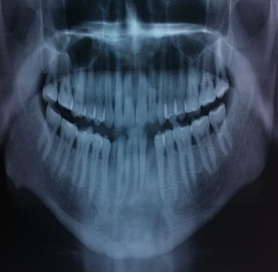
Figure 1: Panoramic radiograph showing radiolucent dentigerous cyst
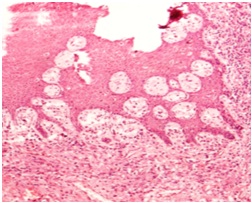
Figure 2: Normal epithelial lining of dentigerous cyst (H&Ex100)
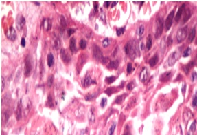
Figure 3: Cyst lining epithelium showing dysplastic changes (H&Ex400)
Discussion
Squamous cell carcinoma arising from dentigerous cyst is a
rare malignant tumor and is exclusively localized in the jaws arising from the
remnants of odontogenic epithelium. Its incidence being 1-2.5% of all
odontogenic tumors [4] and 1-2% of all intraoral cancers [5]
and the commonest site is third mandibular molar followed by canine maxillary
teeth. The age group involved is 37-90 years with male preponderance and M:F of
3:1[6].
The differential diagnosis includes
unicystic ameloblastoma, invasion of cyst from primary carcinoma of jaw and
cystic change in a primary carcinoma of the jaw. Unicystic ameloblastoma
presents as a painless slow growing swelling showing soap bubble radiolucencies
on radiology and a well developed stellate reticulum above basal layer with
reverse polarity. Squamous differentiation in these ameloblastic nests of cells
is well known but they usually lack cellular pleomorphism and mitotic activity,
while retaining the peripheral palisading of their columnar basal cells and the
loose central stellate reticulum. Invasion of cyst from primary carcinoma of jaw
presents as ulceroproliferative lesion in gingival/labial mucosa showing
radiolytic lesions on X-ray and large nest of squamous cells with no evidence of
dysplasia in epithelial lining of the cyst. Cystic change in a primary carcinoma
of the jaw presents as ulceroproliferative lesion in oral cavity showing
osteolytic lesions on X-ray and nests of squamous cells with cyst macrophages.
The present case showed nests of malignant squamous cells with dysplasia in
epithelial lining of the overlying cyst wall. No stellate reticulum was
identified in the nests and there was no peripheral palisading as well, thus
ruling out squamous differentiation in an ameloblastoma (Figure 4).
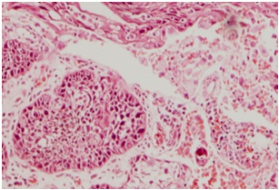
Figure 4: Nest of malignant squamous cells. Note absence of central stellate reticulum (H&Ex100)
Conclusion
Development of SCC from residual cyst is rare, however should always be considered in differential diagnosis of squamous cell carcinomas in jaw region. Hence, proper and adequate biopsy from the representative area and careful histopathological examination scores utmost importance. It is imperative that the following diagnostic criteria should be fulfilled to give an accurate diagnosis: a) histopathological confirmation of SCC b) transition of cyst lining epithelium from normal to dysplastic to malignant c) absence of central stellate cells in nests of neoplastic squamous epithelial cells d) tumor not involving the oral mucosa e) exclusion of metastasis from any other primary site.
Key Message
It is very important to be clinically aware of the significance of the malignant potential of an apparently innocuous cystic lesion.
Author’s Contribution
SS: Contributed to the pathological diagnosis and edited the
final manuscript.
MB: Literature search and drafting of manuscript.
SS:
Literature search and interpretation of results.
N: Helped in drafting the
manuscript.
PM: Treated patient & provided clinical details.
Conflict of Interests
The authors declare that there are no conflicts of interests.
Ethical Considerations
Written informed consent was obtained from the patient for
publication of this case report.
Funding
None Declared
Acknowledgement
None
References
[1].Bradley N, Thomas DM,Antoniades K,Anavi Y-Squamous cell carcinoma arising in an odontogenic cyst. Int J Oral Maxillofac Surg 1993;22:260-263. [pubmed]
[2].Yoshida H, Onizawa k,Yusa H-Squamous cell carcinoma arising in association with an orthokeratinized odontogenic keratocyst:report of a case. J Oral Maxillofac Surg 1996:54:647-651.[pubmed]
[3].Gardner A: The odontogenic cyst as a potential carcinoma: a clinicopathologic appraisal. J Am Dent Assoc 1969;78:746-755.[pubmed]
[4].Bradley N, Thomas DM,Antoniades K,Anavi Y-Squamous cell carcinoma arising in an odontogenic cyst. Int J Oral Maxillofac Surg 1993;22:260-263.[pubmed]
[5].Matsuzaki H,Karase N,MatsumuraT,et al. Solid-type inraosseeous squamous cell carcinoma of the mandible: a case report with histopathological and imaging features. Oral Maxillofac Radiol 2012;114:71-77.[pubmed]
[6].Van der Waal I, RauhamaaR,van der Kwast W A,et al. Squamous cell carcinoma arising in the lining of odontogenic cysts. Report of 5 cases. Int Oral Surg 1985; 14:146-152.[pubmed]
[7].Johnson LM, Sapp JP, Mcintire DN: Squamous cell carcinoma arising in a dentigerous cyst. J Oral Maxillofac Surg 1994;52: 987-990. [pubmed]

