Case Report

Taenia saginata anchored to the duodenum presenting with haematemsis in a healthy adult male
*Mohamed Osman Mohamed Suliman,
Associate professor of surgery, M. S. Idris GIT bleeding Centre, Ibn Sina specialist Hospital, Khartoum, Sudan.
- *Department of surgery, Faculty of medicine, University of Bahry. Head department of surgery, Nile College.
- Submitted: Monday, November 24, 2014
- Accepted: Sunday, February 15, 2015
- Published: Sunday, February 22, 2015
This is an Open Access article distributed under the terms of the Creative Commons Attribution License (http://creativecommons.org/licenses/by/3.0), which permits unrestricted use, distribution, and reproduction in any medium, provided the original work is properly cited
Abstract
Introduction
Taenia saginata is found all over the world, for example; Sudan, Great Britain, China, Iran and Indonesia. It infects people with poor hygiene.
Finding
This case report is about the finding of a tape worm (Taenia saginata) anchored to the mucosa of the second part of the duodenum in a healthy adult male. It presented with haematemsis for which oseophago-gastro-duodenscopy was done. No part of the worm was seen by the patient in the stools.
Conclusion
The worm was removed by the scope and treatment was given to the patient who remained well on follow up.
Keywords
Tape worm in duodenum; Tape worm in stomach; Haematemsis; Gastroduodenoscopy
Introduction
Infection with Taenia saginata occurs in all parts of the world including Britain, Sudan, Cina, Iran and Indonesia
[1]. No significant symptoms are caused by this worm a part from mild, vaque discomfort. Usually parts of the tape worm are passed with the stools and this is considered to be the commonest symptom. This is because the worm has very little contact with the mucosa without causing any immunological response from the body [2].
Case Report
A thirty one year old male, who is a laborer from a low socio-economic class, was admitted to M S Idris GIT bleeding centre in Khartoum, Sudan complaining of haematemesis for one day. The amount of haematemsis was small, the blood was red with no clots and he vomited once. There was no malena. This episode of haematemesis was preceded by nausea and epigastric discomfort for about a month for which the patient did not see a doctor and took no treatment. The patient did not notice any parts of the worm in his stools. He had no other health problems.
On examination he looked well, not anemic or jaundiced. His abdomen looked and felt normal with no epigastric guarding or tenderness. The labrotary investigations were all normal, including stool general which was done after the endoscopy.
Upper GIT endoscopy was done. No inflammatory or neoplastic lesion was found but a long, white object was seen extending from the stomach to the duodenum. On close scrutiny it was recoqnized as a tape worm. The head was gripping the duodenal mucosa of the second part firmly (Figure 1). The body ran through the pyloric canal, which was kept open, up to the proximal part of the stomach (Figure 2). The worm was identified in the labrotary as
Taenia saginata.
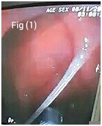
Figure 1: Taenia saginata anchored to the duodenal mucosa.
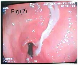
Figure 2: Taenia saginata seen extending from the open pylorus to the stomach.
Discussion
Rarity of this finding
Only three reports were detected in the literature by searching the internet in which this worm was found in the stomach and duodenum. Only in one of these cases [3] there was atrophic gastritis, but in the present case and the other cases there was no atrophy of the gastric mucosa. This implies that the worm must be resistance to gastric juice digestion.
Comparison of presentation between the cases
One of the cases presented with haematemesis and malena [3]. A female patient of 80 years was complaining of epigastric pain only. The second patient was also a female of 54-years, complaining of dyspepsia, epigastric pain, nausea and vomiting on and off for two months
[4]. The third was a 60 year female patient presented with haematemesis and malena
[5] and again one year later she presented with epigastric discomfort and no bleeding. In both presentations she was found to have taenia in the stomach. All these three patients had normal findings on clinical examination a part from anaemia.
A problem in removal
In the present case the worm was held by a biopsy forceps near its head and pulled to release it from the duodenal wall but it was cut. Again the head was held and removed with the rest of the worm.
Conclusion
The patient in this case report was given praziquantel, 10 mg/kg. Stool analysis and endoscopy follow up after one month was normal.
Learning points
-
Taenia saginata can reside in the stomach and duodenum inspite of the digestive enzymes.
-
It can present with haematemsis and/or malena or other upper gastro-intestinal symptoms.
-
Passage of parts of the worm with stools may not occur even if the worm is very long.
Consent
The written informed consent was obtained from the patient
and is available with authors.
Competing interests
The author declares that there is no competing or conflict of interest.
Author contribution
The author did the endoscopy, took care of the patient and followed him up,
the author concieved the idea and prepared the manuscript.
Acknowledgement
Thanks to Dr. Isam Ali Ahmed who took the film.
References:
[1]- John Macleod, editor. Davidson,s Principles and practice of Medicine. 14th ed. Churchill Livingstone; 1984, p 788.
[2]http://bioweb.uwlax.edu/bio203/s2009/temanson_caro/Taeniasis.htm
[3]İsmail Hakkı Kalkan, Aydın Şeref Köksal, Erkin Öztaş, Ayşe Yasemin Tezer Tekçe, Hakan Yildiz, Öykü Tayfur, Endoscopic removal of an immigrant (Taenia saginata) from the stomach of a geriatric patient. Geriatrics &Gerontology International, 2013; 13: 232–233.
[4]Mohsen Masoudi, Zahedin Kheyri and Ali Ali-Asgari , Photo Quiz: Tapeworm in Stomach, Archives of Clinical Infectious Diseases . July 2012; 7(3): 104-5.
[5]Wen-Shen Liao, Ming Jong Bair, Taenia in the Gastrointestinal Tract. N Engl J Med. Sep 2007;357: 1028 [Pubmed]


